
Equipments
Optical Coherence Tomography (OCT):
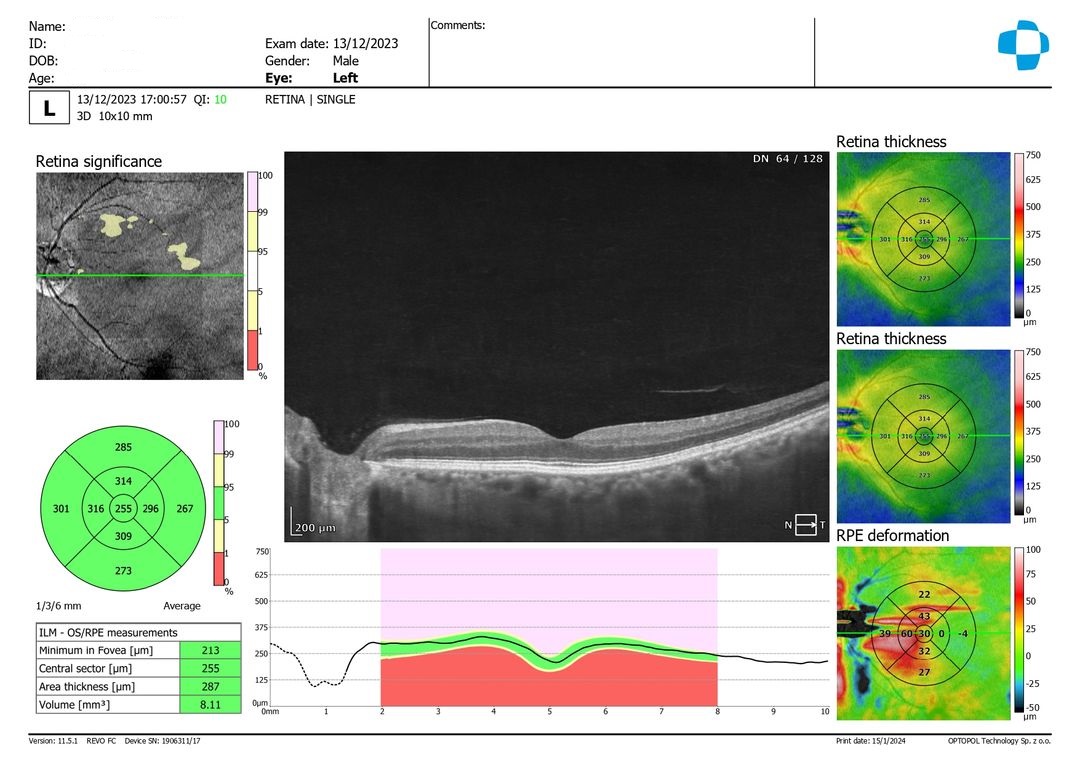
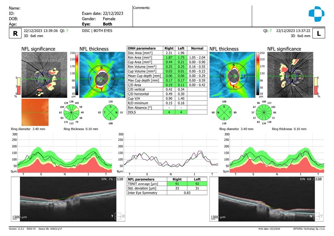
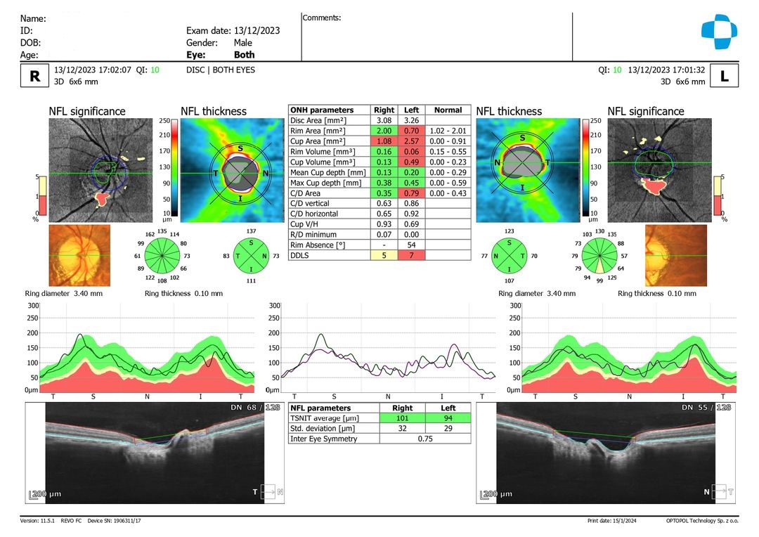
Optical Coherence Tomography (OCT) is a non-invasive imaging test that uses light waves to take cross-section pictures of your eye from the cornea to the retina. It provides richly detailed 3D images with high resolution, allowing for more precise examinations of key structures in the eye such as the cornea, optic nerve disc, and macula. This aids in the effective detection and assessment of serious eye conditions at an early stage.
Main Functions:
- Scans and visualizes the optic nerve structure, aiding in the diagnosis of glaucoma, optic nerve pathology, and monitoring disease progression.
- Scans and visualizes the macular structure, crucial for diagnosing age-related macular degeneration, diabetic retinopathy, macular edema, and other macular diseases, as well as tracking disease progression.
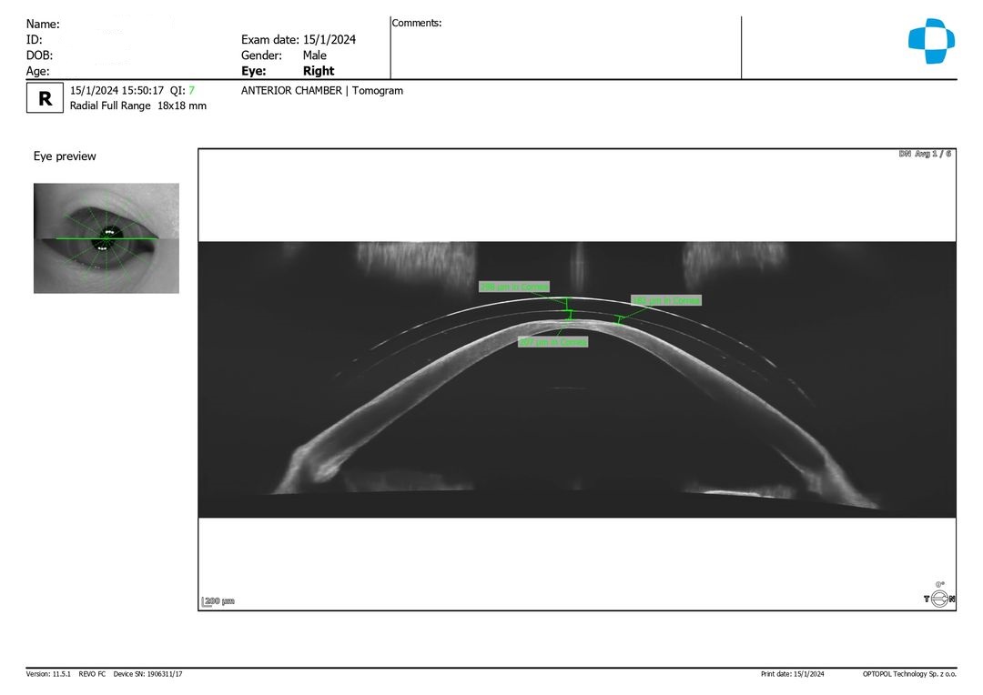
- Corneal and anterior chamber examinations can determine corneal thickness and help identify individuals at high risk for acute glaucoma.
- Scleral lens fitting involves precise detection of lens performance on the eye after settling, providing detailed imaging to patients for improved communication and fitting experience.
- Measurement of axial eye length, useful for monitoring the progression of myopia.
- Glaucoma assessment.
- Macular disease evaluation.
Individuals at Higher Risk Who Should Consider Regular Macular Disease Evaluations:
- Those with a family history of macular disease.
- Patients already diagnosed with macular degeneration or deterioration.
- Individuals experiencing distorted or wavy vision.
- Those with high degrees of myopia (nearsightedness).
- Long-term diabetics.
ESP Eye Surface Profiler


Berlin Optical is the first optometric center in East Asia to introduce this cutting-edge technology, the Eye Surface Profiler (ESP). This device is currently the most precise scleral topography instrument globally, featuring an unprecedented 20mm diameter scanning area and 500,000 measurement points. Its revolutionary analytical technology not only provides corneal profile analysis but also includes profiling of the entire corneal edge and sclera. Additionally, it can accurately map the 3D topography of the cornea and sclera, generating a 3D “height map” of the eye surface to precisely customize the most suitable scleral lenses for each client.
💡 Scleral lenses are FDA-cleared specialty contact lenses used during the day to treat ocular surface diseases such as dry eye syndrome and Sjögren’s syndrome. They can also correct vision for patients with high astigmatism, keratoconus, and post-refractive surgery regression. The unique “tear lens” feature allows wearers to achieve clearer vision than with traditional glasses or contact lenses.
Traditionally, fitting scleral lenses required prolonged trial fittings and continuous modifications to perfect lens fitting. Now, with the ESP, we can scan the scleral topography and 3D map. We then combine this data with the results from trial fittings to send to our US manufacturer for personalized scleral lens customization. This ensures that the lens curvature perfectly matches each client’s unique scleral shape, significantly enhancing comfort and accuracy. The complex fitting process has been simplified into just four easy steps for clients:


Main Functions:
- Examines the topography of the cornea and sclera (the white part of the eye) to create a three-dimensional map. Data is directly transmitted to overseas manufacturers for custom-made specialty contact lenses, including scleral lenses and orthokeratology (OK) lenses.
- Overcomes the drawbacks of traditional scleral lens fitting methods, which require prolonged trial fittings and multiple adjustments, enhancing the precision and efficiency of fitting scleral lenses and other specialty contact lenses
OVITZ xwave Aberrometer

Traditional eyeglasses or contact lenses can only correct Lower-Order Aberrations (LOA), such as myopia (nearsightedness), hyperopia (farsightedness), and astigmatism. However, they are unable to address Higher-Order Aberrations (HOA), such as coma, trefoil, and spherical aberration. These higher-order aberrations are often the primary cause of issues like ghosting (double vision), glare, poor night vision, and starbursts. Scleral lenses with standard optics utilize the tear reservoir formed between the lens and the cornea to correct irregular astigmatism and higher-order aberrations originating from the corneal surface. Nevertheless, they cannot neutralize higher-order aberrations coming from deeper corneal layers or the internal structures of the eye. These uncorrected optical imperfections mean that some patients, even after achieving visual acuity of 1.0 (20/20) or better with standard scleral lenses, still experience "blurry vision" or a lack of clarity.
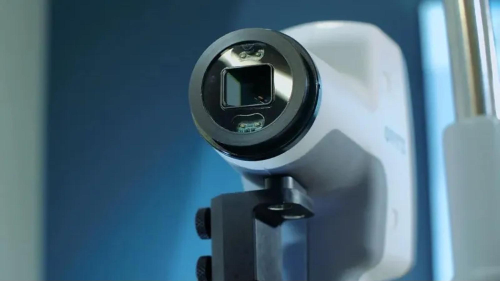
The OVITZ xwave is the world's first aberrometer specifically designed for scleral lenses. This advanced aberrometer precisely captures the total HOA distribution of the eye, and sends the details to the scleral lens manufacturers in Europe and the United States. These manufacturers then engrave the corresponding optics onto the finished scleral lenses to counteract the wearer's optical imperfections. This higher-order aberration customization, also known as wavefront-guided technology, is also used in high-end refractive surgery and cataract surgery to enhance postoperative visual outcomes.
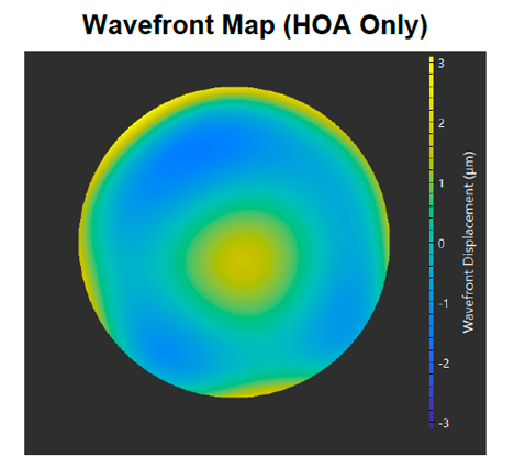
A wavefront map generated by the OVITZ xwave system. If incoming light is perfectly focused inside the eye, the wavefront displacement value will be zero. Areas where greater aberrations occur are displayed in warmer or cooler color tones. The larger the deviation, the more noticeable optical imperfections such as double vision or glare become.
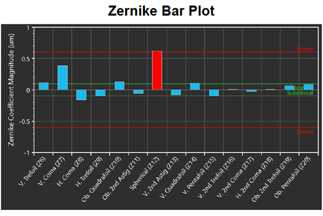
OVITZ xwave’s total higher-order aberration analysis display. The analysis reveals that the patient’s visual quality is significantly affected by pronounced fourth-order spherical aberration.
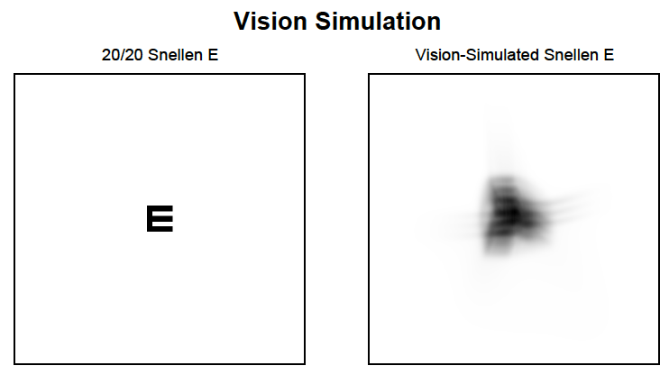
After scanning the total-eye aberrations, the instrument simulates the patient’s affected visual quality.
Berlin Optical is proud to be the first and currently the only advanced scleral lens fitting center in Asia to introduce this HOA customization technology. We are also the only center in Asia that is also equipped with the ESP Scleral Topographer. Our world-class instrumentation, combined with professional service, allows us to craft fully personalized scleral lenses with exceptional vision and comfort.
Primary Functions:
- To investigate the reasons why patients still experience glare and poor night vision even after wearing glasses or standard contact lenses.
- To manufacture wavefront-guided scleral lenses, addressing the issue where some patients have good measurable vision (visual acuity) but still suffer from poor visual quality.
Corneal Topography


Corneal topography is a non-invasive medical imaging technique that provides a detailed analysis of the cornea's shape and curvature. This analysis is crucial for diagnosing various corneal conditions. It is performed simply by looking into the device as instructed by an optometrist.
Main Functions:
- Examines the corneal topography and tracks changes over time, assisting in the diagnosis of conditions like keratoconus, assessing astigmatism, and post-surgical recovery.
- Collects corneal curvature, diameter, and other data, useful for fitting orthokeratology lenses, scleral lenses, and other specialized contact lenses, as well as tracking lens effectiveness.
- Evaluates tear film quality, aiding in the diagnosis of dry eye syndrome.
Slit Lamp


A slit lamp is a specialized microscope used in eye examinations that provides a high-intensity focused light source for detailed observation of all eye structures, from the cornea and iris to the lens and retina. A slit lamp exam is a non-invasive diagnostic process that aids eye doctors and optometrists in detecting and diagnosing various eye diseases, such as cataracts, glaucoma, and corneal damage or diseases.
Main Functions:
- Comprehensive examination of the external and internal conditions and health of the eye, used to diagnose diseases from the eyelids, eyelashes, cornea, conjunctiva, lens, and retina, such as dry eye syndrome, cataracts, glaucoma, and macular diseases.
- Equipped with digital photography capabilities to record the current state of the eye, facilitating monitoring of changes.
Digital Retinal Photography


Digital retinal photography is a key ophthalmic imaging technique that examines and records the current state of the retina. It is highly useful for diagnosing various retinal diseases, including glaucoma, macular degeneration, and diabetic retinopathy.
Automatic Computerized Refractometer

An automatic computerized refractometer can initially estimate a patient's prescription and measure the corneal curvature. Newer models are further equipped with non-contact tonometry and corneal thickness measurements, playing an important role in glaucoma screening.
Photo keywords: Nidek Tonoref III, non-contact tonometry, glaucoma screening.
Digital Lens Fitting System

Equipped with the advanced French Essilor VISIOFFICE® 3 digital optometric measuring system, this system uses an HD color camera that can precisely capture a range of eyewear and eye interface parameters including pupillary distance, fitting heights, eye rotation center, pantoscopic tilt, and frame wrap angle with just two simple photos. Its accuracy reaches up to 0.1 millimeter and 0.1 degree and provides a 3D reconstruction of eye anatomy, allowing for lenses and coatings to be customized based on an individual's lifestyle and natural posture.
This system not only observes personalized data such as dominant eye, reading habits, and posture but also converts collected eye data into 3D images for easier explanation of eye conditions to clients. The user-friendly software allows clients to compare different frames and lens options, enhancing the visual experience. Furthermore, this system adheres to social distancing protocols, offering eye care professionals a safe and innovative option to enhance service quality and expertise, providing clients with a unique and comfortable eyewear fitting experience.
.svg)
.svg)
.svg)
Service

